What are organ-on-chips?
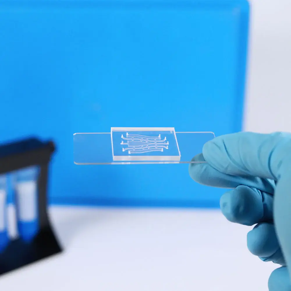
Organ-on-chips (OoC) or Micro-Physiological Systems (MPS) are microfluidic systems that allow the growth of cells to model biological tissues. These platforms combine a multicellular environment with a 2D or 3D structure and mechanical cues to mimic the physiological environment of an organ.
Why is 3D important in cell-culture?
Currently, in vitro testing occurs mainly on a single cell type, cultured in 2D, in hard, flat, polystyrene flasks or plates. Even if those techniques have been useful in deciphering some phenomena, it is clear that they do not reproduce the environment of cells inside a body. In the human body, organs are made of multiple cell types, arranged in a 3D structure, and surrounded by extracellular matrix and other tissues that are usually a lot softer than plastics. In 2D monolayers, cell morphology, and polarity are forced and not representative of reality. In addition, the constraints in terms of nutrient access and spreading or migration are non-existent in 2D and again do not model what happens inside the body. Cellular interactions with the environment or with other cell types are also lacking in 2D models.
Why are mechanical cues important in cell development?
As exposed above, in the body, tissues are exposed to different types of mechanical forces from their environment. Some organs like muscles are subjected to compression and stretch cycles that contribute to cell development and fate. Without those mechanical forces, the cell would behave differently, thus it is important to reproduce those forces in a 3D in vitro model to better mimic pathologies and also take this natural motion into account for therapy development. Not only should one take into account the nature of the biomechanics stimulation but also its amplitude, frequency, and duration.
Blood circulation is another component of the biomechanics in the body. The shear stress induced by the flow inside capillaries, vessels, and arteries has a profound impact on endothelial cells and overall tissue remodeling. Thus, it is crucial to reproduce a type of blood flow within those new models for mechanical reasons but also biological reasons. Indeed, blood carries nutrients to tissues and cells, and having a dynamic flow allows for a constant supply of fresh nutrients as opposed to a static culture where the same media can stay for several days. Similarly, cellular metabolic waste will be continuously removed and will not accumulate in the cell culture, which could induce toxicity in the cells.
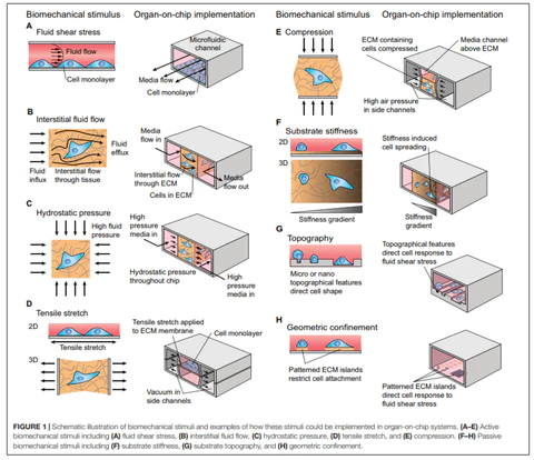
Thompson CL, Fu S, Heywood HK, Knight MM and Thorpe SD (2020) Mechanical Stimulation: A Crucial Element of Organ-on-Chip Models. Front. Bioeng. Biotechnol. 8:602646. doi: 10.3389/fbioe.2020.602646
Why are organ-on-chip developed?
The need for those tools in research emerged in the drug discovery process to address several unmet expectations.
- Decrease costs and duration: currently it can take up to 20 years and billions of dollars to get a drug on the market
- Minimizing animal experimentation: pre-clinal testing of toxicity and efficacy of drug candidates is performed on animals such as rodents and primates. Each year, millions of animals are used to model human biology which poses both ethics and scientific relevance considerations.
- Get more relevant models of human biology: there is a need for a systemic human model to test toxicity and efficacy in more physiological conditions than current human in vitro or animal in vivo models.
There is high hope that the development and use of new 3D human models might help increase the success rate of drug testing while reducing the costs and duration of drug development.
Which applications can organ-on-chip be used for?
Organ-on-chip can be used for a variety of applications all along the drug development process, including the critical study of the blood-brain barrier. They can be used to model diseases to decipher some mechanisms, but also to test drug candidates’ safety and metabolism. An ultimate goal of organ-on-chip development would be to use them for personalized medicine, using the patient’s specific cells on which one could try several molecules to choose the most effective treatment for the patient. Overall, for optimal use and adhesion of researchers to organ-on-chip, models should be developed to be associated with classic research techniques such as high-resolution imaging, RNA and protein expression, or cell-secreted biomarkers measurement.
References:
- https://www.upmbiomedicals.com/resource-center/learning-center/what-is-3d-cell-culture/2d-versus-3d-cell-culture/
- Franzen N, van Harten WH, Retèl VP, Loskill P, van den Eijnden-van Raaij J, IJzerman M. Impact of organ-on-a-chip technology on pharmaceutical R&D costs. Drug Discov Today. 2019 Sep;24(9):1720-1724. doi: 10.1016/j.drudis.2019.06.003. Epub 2019 Jun 8. PMID: 31185290.
- Thompson CL, Fu S, Heywood HK, Knight MM and Thorpe SD (2020) Mechanical Stimulation: A Crucial Element of Organ-on-Chip Models. Front. Bioeng. Biotechnol. 8:602646. doi: 10.3389/fbioe.2020.602646

3D Cell Culture: Advantages and Disadvantages
[…] Organ-on-chips, displaying several connected chambers/channels to co-culture different types of cells, oopened the way towards the study of organ cross-talks, secretory functions and dynamic cell cultivation; […]