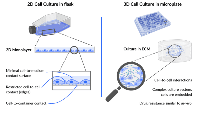
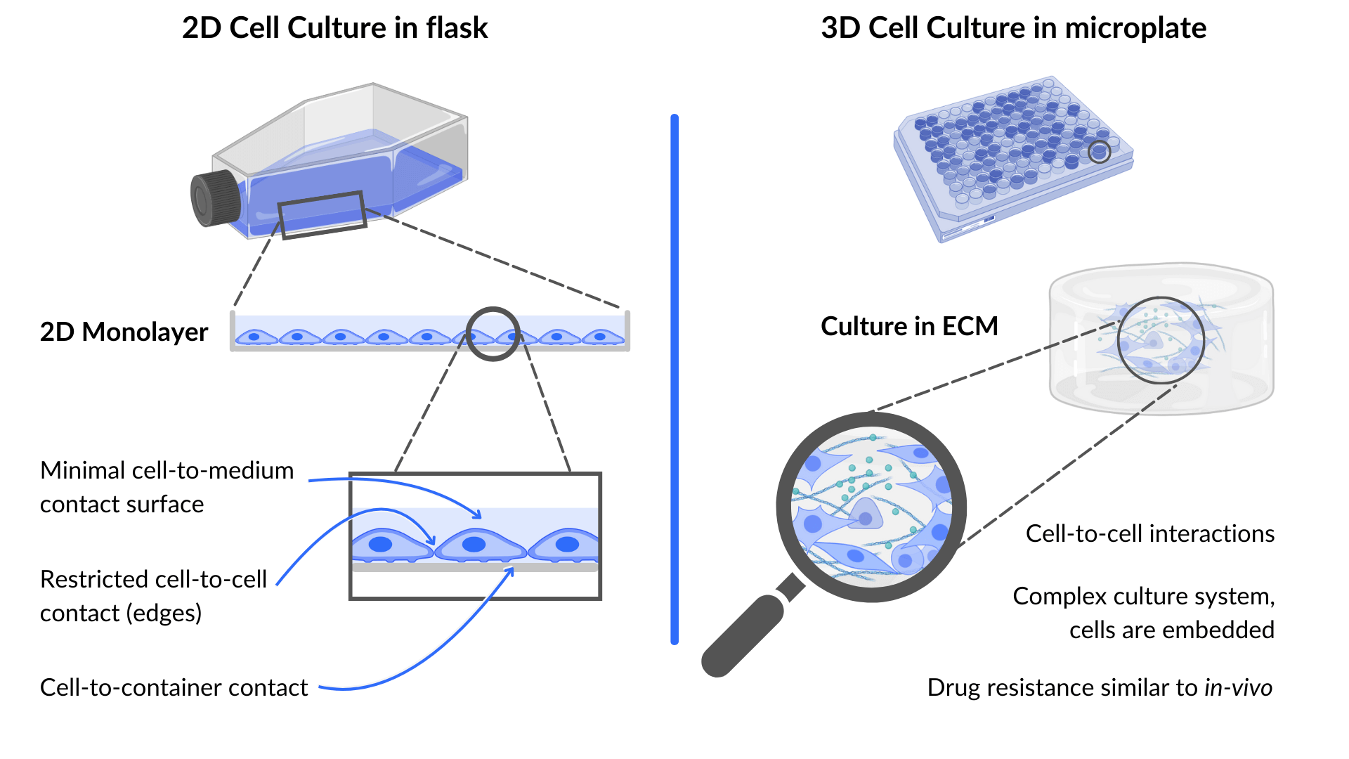
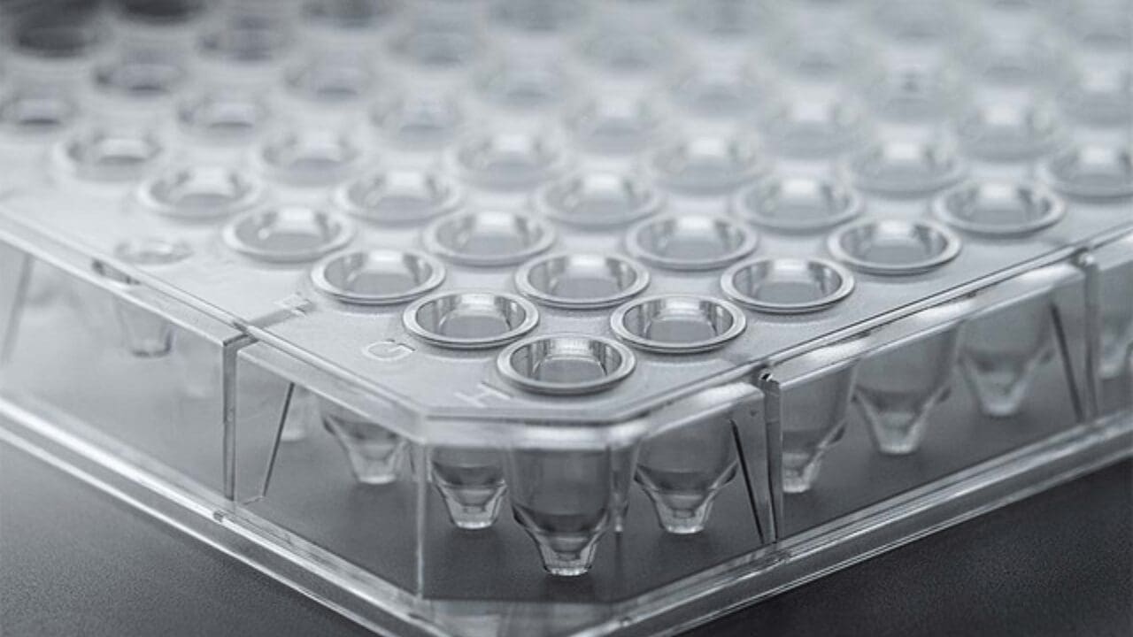
Cell culture refers to all laboratory techniques enabling the growth of cells in an artificial environment, under physiological controlled conditions. In vitro research has for decades been associated with 2D monolayers and cell culture flasks. While this allowed significant advances in many fields from cellular mechanisms understanding to drug development, it presents important limitations. The first being that our body is not organized in 2D. In response to these limitations, scientists have developed innovative approaches to cultivate cells in three dimensions. The past decade has brought its share of innovations in biotechnologies to move from 2D to 3D cell culture such as spheroids microplates or bioprinting.
Here we will discuss what 3D cell culture is and how its advantages and limitations can impact many fields of research.
Table of Contents
3D cell culture - Definition and Application
3D cell culture has been defined as “a cell culture that can mimic a living organ’s organization and microarchitecture” (4. Huh et al., 2011). In other words, it is the ability to maintain cells in artificial spaces where they can grow and interact with their environment in all three dimensions. This allows better mimicking of the real environment, i.e. the human body, of the studied organs. Additionally, this artificial environment can be tuned in terms of shape, material or even chemical composition to mimic this in vivo environment as physiologically as possible.
‘3D cell culture’ is a general term used to describe several techniques and methods:

The use of scaffolds, whether they are synthetic (made of ceramics or polymers for example) or natural like extra-cellular components and hydrogels to recreate the environment of an organ both mechanically and biologically;

Bioreactors, allowing large-scale cultivation of many types of cells, in a highly controlled environment and precise regulation of culture parameters;

Organ-on-chips, displaying several connected chambers/channels to co-culture different types of cells, oopened the way towards the study of organ cross-talks, secretory functions and dynamic cell cultivation;

Organoids, which replicate the 3D architecture of the different types of cells composing an organ, and allowing better understanding of organ development and regeneration as well as the study of tumors.

Bioprinting, using bioinks to recreate 3D structures, from scaffolds, to organs.
Historically, the need for 3D in vitro models grew with the limitations on animal use in research and the amount of national or international directives to develop non-animal based methods for pharmaceutical but also cosmetic research. These limitations came from ethical (suffering caused to the animals) and biological (lack of translation from animals to humans) arguments.
3D cell culture can be used in all applications 2D cell culture is used and even more as it provides an additional degree of complexity to gain a deeper understanding of cell behavior and mechanisms. The applications include: drug discovery and drug metabolism, tissue development, cancer research and many others.
Why is 3D cell culture better than 2D cell culture?
The desire and need to implement 3D cell culture techniques in many research projects has been growing over the last years because of the numerous advantages associated with this innovation. The possibility to grow in all three dimensions and interact with a physiological environment has indeed major positive implications for cells as compared to growing in 2D, on a rigid plastic surface.
In 3D cell cultures:
- Cell morphology is more natural and resembles more closely that of in vivo environment;
- Cell spreading is sterically constrained and cellular proliferation is slowed down in comparison to 2D where the proliferation rate is much higher than what’s observed in vivo
- Natural cell polarity is established, ensuring a more physiological behavior of the cells.
- More realistic cell-cell and cell-matrix interactions are promoted, replicating more physiologically exchange of mechanical, biological and chemical signals.
- The access to nutrients, including oxygen is also modified and more realistic in 3D cultures as cells are exposed to spatiotemporal gradients and not all cells are exposed to the same amount of each nutrient at the same time as it is the case in 2D.
In summary, cells cultivated in 3D have much more similarities with in vivo cells in terms of structure and thus in terms of functions. It has been shown that all the parameters listed above are important regulators of gene expression patterns. Thus, with a more physiological environment, cells should also behave more physiologically than in 2D cell culture. It is expected that the observed effect of drugs and molecules on these models would be closer to what happens in the body, enhancing the predictability of efficacy and toxicity of newly developed drugs and improving the translation success from preclinical to clinical studies.
What are the problems with 3D cell culture?
Despite the numerous advantages of 3D cell culture discussed above, it also comes with some limitations, which can be overcome but are important to have in mind when thinking of transitioning from 2D to 3D cell culture.
- Because of the increased complexity inherent to the 3D structure, setting up those models usually requires more expertise and training, as well as technicity. It can be at first challenging and time-consuming to find all the right elements and experimental conditions and transition to new protocols and techniques.
- At the moment, as this is a whole new field, there is a global lack of standardization and common protocols. It can be hard to find guidelines for beginners and to easily reproduce and compare results. This standardization question is however at the center of many discussions with key actors of the field.
- Overall, 3D models are more expensive to set up and to maintain. These models may require switching to new laboratory consumables, like hydrogels, scaffolds and plasticware and eventually equipment, especially regarding imaging.
- Imaging indeed can be tricky as it is harder to see cells inside of a 3D structure than on a monolayer. Adding a dimension in image acquisition can thus require expertise, training and potential investment in appropriate equipment.
- In comparison to classic culture flasks used in 2D culture containing millions of cells, scaling up of newly developed 3D models for large-scale studies can be a challenge because of the complexity of the models and the current platforms available.

In the end, 3D cell culture seems to be the logical next step for all researchers trying to decipher the mechanisms behind organ development, biological processes, disease pathophysiology or drug efficacy or toxicology. The field is moving, fast, and many technologies are being developed to help researchers joining the movement towards 3D models.
Sources :
- Cacciamali A, Villa R, Dotti S. 3D Cell Cultures: Evolution of an Ancient Tool for New Applications. Front Physiol. 2022 Jul 22;13:836480. doi: 10.3389/fphys.2022.836480. PMID: 35936888; PMCID: PMC9353320.
- Jensen C, Teng Y. Is It Time to Start Transitioning From 2D to 3D Cell Culture? Front Mol Biosci. 2020 Mar 6;7:33. doi: 10.3389/fmolb.2020.00033. PMID: 32211418; PMCID: PMC7067892.
- https://www.upmbiomedicals.com/resource-center/learning-center/what-is-3d-cell-culture/2d-versus-3d-cell-culture/
- Huh D, Hamilton GA, Ingber DE. From 3D cell culture to organs-on-chips. Trends Cell Biol. 2011 Dec;21(12):745-54. doi: 10.1016/j.tcb.2011.09.005. Epub 2011 Oct 25. PMID: 22033488; PMCID: PMC4386065.


Spheroids and 3D Organoids Cell Cultures: The Main Differences
[…] In the dynamic landscape of biomedical research, the advent of three-dimensional (3D) cell cultures has revolutionized our ability to study complex biological systems outside the human body. Unlike traditional two-dimensional (2D) cultures, which oversimplify cellular interactions, 3D cultures like spheroids and organoids more accurately mimic the intricate structure and function of tissues and organs. This advancement is crucial for gaining deeper insights into fundamental biological processes, disease mechanisms, and drug responses in a more physiologically relevant context. In the past decade, scientists have developed innovative solutions to cultivate cells in 3D. […]