Spheroids, in biology, are 3D aggregates of cells mimicking the architecture and function of tissues. In environments preventing attachment to surfaces, cells can indeed self-assemble into sphere-like structures, called spheroids. There has been growing interest for these structures in the past years as they constitute a 3D model, closer to the in vivo microenvironment of cells in a living organism than the classic 2D cell culture. 3D cell culture displays many advantages for medical research like creating more predictive models to support the development of new therapies. In this blog article, we will explore the basics of cellular spheroids formation and dive into its many applications.
Spheroid Formation - How do cells aggregate?
There are several ways to form spheroids but the biological processes behind this cellular clustering is quite complex and involves several cellular mechanisms. It indeed requires cell-cell adhesion from neighboring cells through transmembrane receptors at the surface of cells called integrins, resulting in the aggregation of cells. This leads to the accumulation of another protein called cadherin, involved in cell-cell adhesion. Then, those assembled cells create tight junctions which enhances the initial cellular adhesion and also increases the spheroid compaction. In addition, complex processes of cytoskeleton remodeling, extracellular matrix (ECM) production and other intracellular signaling occur during the process of spheroid formation.
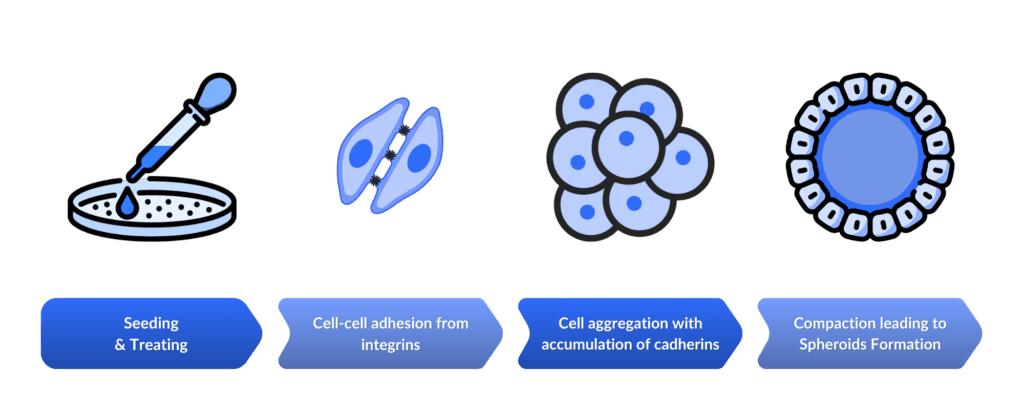
After the initial formation, the spheroids can continue to grow, especially tumor spheroids, and can be kept in culture for weeks. They can be exposed to treatments and other experimental conditions to which they will react by cellular architecture modification, molecule secretion or other cellular behavior. Over time, a gradient of nutrients and oxygen will appear between the internal layers of cells and the external ones. Regular cellular processes may undergo some modifications. For example, cell proliferation rate may be impacted in core cells, less exposed to nutrients and oxygen leading to apoptosis (programmed cell death) or necrosis (uncontrolled cell death).
What are the Techniques to form Cellular Spheroids?
Different cell types have different propensities to form spheroids, hence the development of several methods to promote cell aggregation. Here is a (non-exhaustive) list of these techniques:
- Some cell types can naturally aggregate, through the expression of cell adhesion molecules. For example, cancer cells often self-aggregate into spheroids, eventually becoming tumors.
- While some cells might be relatively prone to form aggregates, they might also benefit from some help from the environment. This help can be brought through the addition of biochemical components promoting cell-cell adhesion, like integrins, cadherins or extra-cellular matrix components and many other molecules.
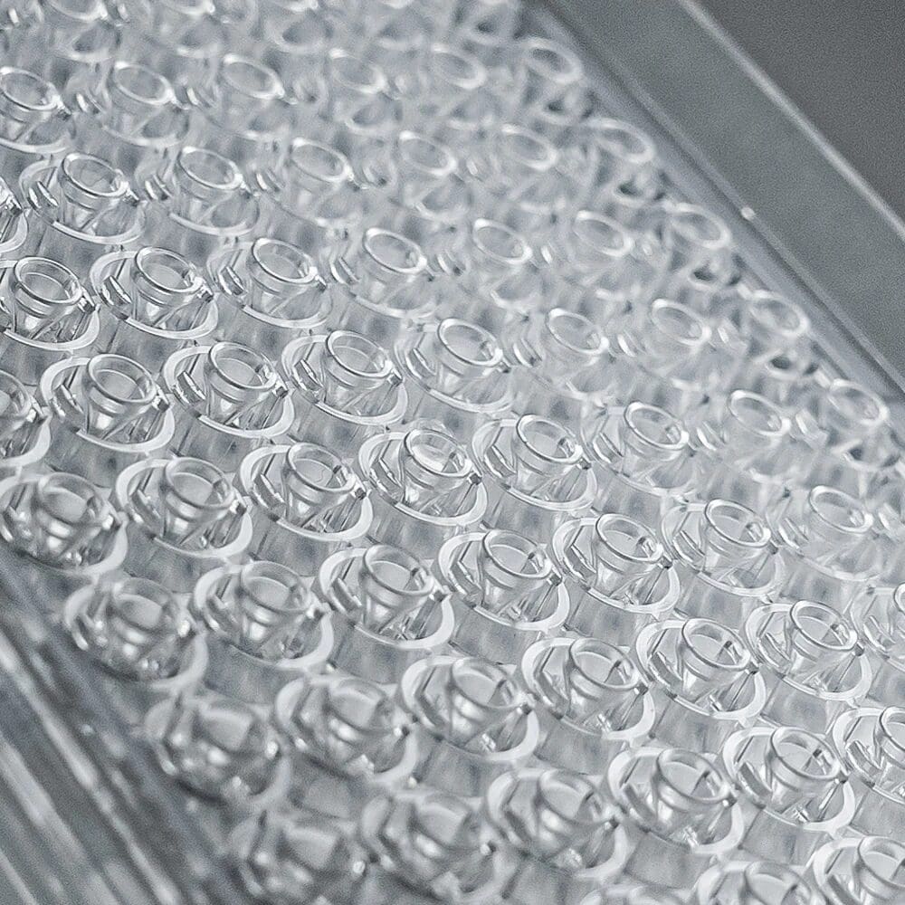
- It is also possible to use low-attachment or non-adherent coatings on the surfaces of the device used for cellular aggregation. In addition, physically constraining the environment might enhance spheroid formation through specific shape of the wells (such as conical wells with special ledges enabling near-complete medium exchange, as seen on the InSphero AKURA microplates) or micropatterning with specific patterns on the surface to guide cellular aggregation.
- Maintaining cells in suspension through agitation can prevent cells from attaching to the surface and promote cell-cell contacts. While this is quite simple to put in place, it often results in high variability of spheroid size.
- Electric and magnetic fields, ultrasound or acoustic waves can also be used to favor cell clustering.
- Microfluidics can also be used to form cell aggregates, with several advantages: using less reagents, displaying a well-controlled environment, improved size homogeneity between spheroids of same and different batches, better modeling and cell viability through fluid dynamics.
- Another very common option is to use the hanging drop method. It leverages gravity to facilitate cell-cell interactions in a suspended droplet of medium. While this can be done on the lid of a Petri dish, some devices were specifically developed to improve the reproducibility and high-throughput production of these hanging drops.
Techniques summary
| Technique | Description |
|---|---|
| Natural Aggregation | Expression of cell adhesion molecules. |
| Biochemical Components | Promote cell-cell adhesion. |
| Low-attachement or non-adherent coatings | Enhance spheroid formation. |
| Constraining Environment | Guiding cell aggregation with specific well shapes or micropatterning. |
| Suspension and Agitation | Prevent surface attachment and promote cell-cell contacts. |
| Physical fields | Favor cell clustering by using electric and magnetic fields, ultrasound, or acoustic waves. |
| Microfluidics | Forming cell aggregates in controlled environments. |
| Hanging Drop | Leveraging gravity to facilitate cell-cell interactions in a suspended droplet of medium. |
What other Factors can influence Cell aggregation?
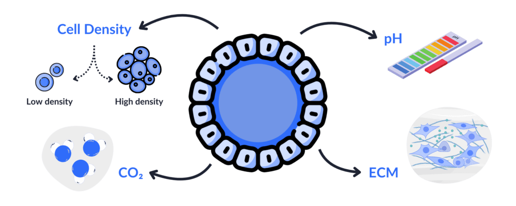
- Cell density has an impact and it is important to determine the optimal number of cells for a given volume which will promote effective cell-cell interactions for successful spheroid formation. If there are not enough cells, there might be insufficient cell-cell contacts leading to poor aggregation. Yet, too many cells can induce poor access to nutrients, cell stress and eventually cell death. Having a constant cell density will also contribute to homogeneous and uniform spheroids for reproducible experiments.
- Cell culture and environmental conditions: all the factors important for cell viability and growth will also impact spheroid formation. Indeed, it is important to have sufficient levels of nutrients and oxygen, optimal temperature, pH and CO2 levels, and an appropriate medium composition for the type of cell chosen. If the conditions are not optimal, this might impact cellular interactions and communications and induce cell stress up to cell death. Metabolites produced by cells within the spheroid also influence other cells. Especially, the metabolites produced by the cells located at the center of the spheroid might not reach the cells of the outer layers, and thus cells will not be exposed to the same metabolites, or concentration of them depending on where they are located in the spheroid. This has an impact on intercellular communication and can also play a role in the different steps of spheroid formation. Mechanical forces and conditions also contribute to spheroid formation whether they are natural or constrained by the method of aggregation chosen.
- Adhesion molecules and extracellular matrix components play an important role in cell adhesion and cellular aggregation. Promoting the expression of these molecules can also be an adjusting parameter.
What are the types of cellular spheroids?
Many types of spheroids exist, each having a unique contribution to research:
Multicellular Tumor Spheroids (MCTS)
Multicellular tumor spheroids (MCTS) are pivotal in modeling solid tumors. These spheroids, intermediates between 2D culture and in vivo tumors, replicate crucial features of tumors, such as the formation of nutrient gradients, and the development of an hypoxic core with less access to oxygen but also physiological intercellular communication and cellular heterogeneity. By closely mimicking the tumor microenvironment, MCTS are essential for studying cancer biology and testing anti-cancer therapies.
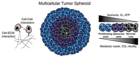
Source: Kamatar, A.; Gunay, G.; Acar, H. Natural and Synthetic Biomaterials for Engineering Multicellular Tumor Spheroids. Polymers 2020, 12, 2506.
Stem Cell Spheroids
Derived from induced pluripotent stem cells (iPSCs), they have the remarkable ability to differentiate into various cell types. This versatility makes them a cornerstone in regenerative medicine and tissue engineering research. These spheroids provide insights into stem cell behavior and potential therapeutic applications.
Embryoid Bodies (EBs)
Included in the previous category, Embryoid Bodies (EBs) are formed from embryonic stem cells (ESCs) and iPSCs. They are used to study early embryonic development. They can also serve as a model for tissue engineering, regenerative medicine and lineage specification.
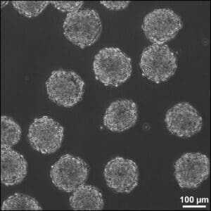
Phase image of embryoid bodies (EBs) in suspension culture after 24 hours of formation. EBs were created from approximately 1000 mouse embryonic stem cells (D3 cell line). Scale bar = 100 µm.
Source: Embryoid body. (2024, June 22). In Wikipedia.
Tissue-Specific Spheroids
These are often co-culture spheroids, displaying multiple cell types to mimic the complexity of tissues as they are usually not composed of a single cell type. These spheroids facilitate the study of cell-cell interactions, tissue engineering, and disease modeling. They provide a more physiologically relevant model by incorporating diverse cell types that interact within a tissue.

Liver Spheroids
Composed of hepatic cells (hepatocytes, non-parenchymal cells, hepatic stellate cells), these spheroids are used to model liver function and diseases, study drug metabolism, and conduct toxicity testing. They can reproduce liver-specific activities, making them a valuable tool for hepatic studies.

Neurospheres
Derived from neural stem cells or progenitor cells, neurospheres are employed to study neurogenesis, neural differentiation, neurodegenerative diseases, and neural tissue engineering. They offer a dynamic model for exploring the complexities of the nervous system.

Cardiospheres
Composed of cardiac progenitor cells, cardiospheres are instrumental in cardiac regeneration research, heart disease models, and cardiotoxicity testing. They mimic the cardiac microenvironment, providing insights into heart tissue repair and disease mechanisms.

Adipospheres
Formed from adipose tissue cells, adipospheres are used to study adipogenesis, obesity, metabolic diseases, and drug testing. They model the behavior and function of adipose tissue, including fat storage and hormone secretion.
Following the examples described above, it is possible to imagine spheroids for all organs of the body, provided that optimal experimental conditions can be determined and comparison studies to existing models and in vivo are performed to validate the relevance of the model.
Immune Spheroids
These aggregates incorporate immune cells, such as lymphocytes and macrophages, to study immune responses, immunotherapy, and tumor-immune interactions. They are essential for understanding how immune cells interact with other cell types and how these interactions influence disease progression and treatment outcomes.
Vascularized Spheroids
They describe spheroids from the above categories containing vascular components or ones which are derived from endothelial cells. They can be used in research on angiogenesis, vascular biology, and drug testing related to blood vessel formation. These spheroids help understand the formation and function of blood vessels, which is crucial for tissue engineering and cancer research. Vascularization is essential for nutrient supply and cell survival and is thus also critical for the development of relevant and physiological spheroid models of different tissue types.
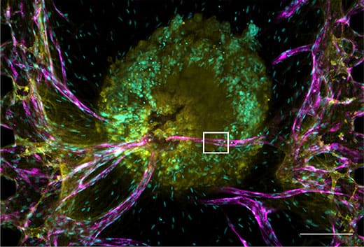
Tumor spheroid and vascular network performed in a microfluidic device.
Source: Nashimoto Y, Okada R, Hanada S, Arima Y, Nishiyama K, Miura T, Yokokawa R. Vascularized cancer on a chip: The effect of perfusion on growth and drug delivery of tumor spheroid.
Biomaterials. 2020 Jan;229:119547.doi: 10.1016/j.biomaterials.2019.119547. Epub 2019 Oct 17. PMID: 31710953.
How can Spheroids be applied in Medical Research?
The development of spheroid cultures has brought a whole new dimension to medical research. These cellular aggregates constitute reliable and cost-effective models to gain insights in disease mechanisms, biological pathways and treatment efficacy and toxicity. They represent a new in vitro model and an alternative to animal testing for better predictability and improved translation to human clinical trials.
| Application | Role |
|---|---|
| Drug Screening | Used for studying both the toxicity and efficacy of newly developed molucules and currently authorized drugs, aiding in drug repurposing. |
| Disease Modeling | Enable a better understanding of disease mechanisms and elucidation of biological pathways. |
| Personnalized Medicine | Potentially used to find the most efficient treatment for individual patients. |
| Cancer Research | Provide a superior in vitro model for tumors compared to 2D cell cultures, aiding in the study of tumor invasion, metastasis, and the development of anticancer therapies. |
| Tissue Engineering | Used as building blocks to develop larger 3D structures when fusioning several spheroids. They can also be used for repairing or replacing injured tissues. |
| Cell Therapy | Employed to deliver cells physiologically in cell therapy developments. |
Many other ways to use spheroids in research and therapy can and will probably be found in the future especially the existing drawbacks of spheroids can be limited.
What are the Limitations of using Cellular Spheroids?
Despite its pivotal role in contemporary biomedical research, spheroid culture still presents some limitations:
📏 Size limitations and reproducibility
The bigger a spheroid is, the more it will be subjected to nutrient gradients and thus to potential cell necrosis at its core. In addition, it is important to develop a production method displaying a good spheroid homogeneity to be able to compare results from different spheroids. It is also important to tend to develop standardized methods in order to get reproducible data.
🤖 Automation and scalability
While many innovations appear in this field, it is important to develop automated systems for the production and study of spheroids in order to make them useful for medical research at a large scale.
🧑🔬 Technical expertise
At first, switching from 2D to 2D may require some laboratory equipment adaptations and technical training.
🔎 Limited analysis
Because of their 3D structure, some endpoints like imaging may be challenging. It might be difficult to access the core of the spheroid and might thus limit the analysis to the outer layers.
🧩 Limited complexity
While more complex than 2D monolayers, spheroids are still less complex in terms of architecture, vascularization, number of cell types, and functions than native tissues.
Conclusion
Cellular spheroids display very promising characteristics for a more physiological, more predictive and more efficient research. Their use in many fields make them a versatile and valuable tool for researchers and medical professionals. The constant innovation in the spheroid field over the last years gives also promising insights towards the potential improvements to come.
Sources :
Białkowska K, Komorowski P, Bryszewska M, Miłowska K. Spheroids as a Type of Three-Dimensional Cell Cultures-Examples of Methods of Preparation and the Most Important Application. Int J Mol Sci. 2020 Aug 28;21(17):6225. doi: 10.3390/ijms21176225. PMID: 32872135; PMCID: PMC7503223.
Decarli MC, Amaral R, Santos DPD, Tofani LB, Katayama E, Rezende RA, Silva JVLD, Swiech K, Suazo CAT, Mota C, Moroni L, Moraes ÂM. Cell spheroids as a versatile research platform: formation mechanisms, high throughput production, characterization and applications. Biofabrication. 2021 Apr 8;13(3). doi: 10.1088/1758-5090/abe6f2. PMID: 33592595.
Lazzari G., Couvreur P. & Mura S. (2017). Multicellular tumor spheroids: a relevant 3D model for the in vitro preclinical investigation of polymer nanomedicines. Polym. Chem. 8, 4947.

